Dermatology Sample CME Question 1

A 62-year-old woman visited the doctor for a black growth on her sole. The patient noted that she had a birthmark in that area of her foot for many years, which was fainter in color and smaller in size, but over the past few months, it had gradually grown and darkened. On dermatological examination, a thin black plaque with a lighter brown rim was seen on the medial sole. Excisional biopsy showed an asymmetric proliferation of severely atypical melanocytes with loss of maturation, pleomorphism, prominent nucleoli, and mitoses. What is the diagnosis?
Can you diagnose the patient? Use the Differential Builder in VisualDx to help you.
- AAgminated nevus
- BAtypical nevus
- CLentigo simplex
- DMelanoma
Dermatology Sample CME Question 2

A 67-year-old man visited the doctor for a cyst-like lesion that developed on his face 3 months prior. The patient reported that the lesion was asymptomatic and caused no discomfort. On examination, a whitish nodule with overlying telangiectasia was seen near the lateral canthus. A thick, cheesy material with a foul odor was expressed upon minimal incision. What is the diagnosis?
Can you diagnose the patient? Use the Differential Builder in VisualDx to help you.
- AApocrine hidrocystoma
- BEccrine hidrocystoma
- CEpidermoid cyst
- DPilar cyst
Dermatology Sample CME Question 3
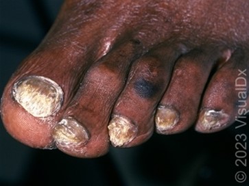
A 62-year-old woman visited her doctor about thick, crumbling, discolored toenails that developed over many months. The patient had a history of diabetes mellitus, peripheral arterial disease, and obesity. On examination, thickened, yellowish nails with subungual hyperkeratosis were seen. The patient appeared well and reported no systemic symptoms. What is the diagnosis?
Can you diagnose the patient? Use the Differential Builder in VisualDx to help you
- ANail psoriasis
- BOnychogryphosis
- COnychomycosis
- DYellow nail syndrome
Dermatology Sample CME Question 4
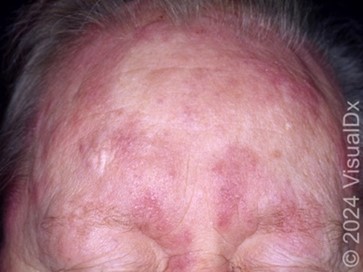
A 71-year-old man visited his doctor about a widespread rash on his upper face and scalp that developed 2-3 months prior. On dermatologic examination, there were widespread deep pink plaques on his forehead and scalp accompanied by patchy hair loss. The patient was not on any medications and had no recent history of trauma or infection. He was otherwise well and had no systemic symptoms. On histopathology examination, mucin deposition in the hair epithelium, stellate-shaped follicular keratinocytes, and perifollicular lymphocytes were seen. What is the diagnosis?
Can you diagnose the patient? Use the Differential Builder in VisualDx to help you.
- AAllergic contact dermatitis
- BAlopecia mucinosa
- CDiffuse mastocytosis
- DTumid lupus erythematosus
Results
Provide your email to see your results and get exclusive discounts on additional board review questions.
VisualDX: Real-World Dermatology Cases + CME Credits
Comprehensive Dermatology Clinical Decision Support, specifically engineered for clinicians to visually improve decision making for optimal patient care. Features expert-curated, real-world case studies and guidance from practicing clinicians. Built by clinicians for clinicians.
Features:
-
Extensive library of dermatological conditions across diverse skin tones
-
Guided differential diagnosis tools for complex cases
- Clinical decision support at point-of-care
-
Educational resources for patient engagement
- Clinicans who use VisualDx save on average 2+ hours a week
CME Opportunities:
-
Earn up to 0.5 AMA PRA Category 1 Credits™ per search
-
CME credits automatically accrue through regular platform use
-
Joint accreditation through the Postgraduate Institute for Medicine
-
Credits available for physicians, pharmacists, and nurses
-
Access CME tracking and certificate printing through your personal account
Trusted by leading institutions and private practices, clinicians rely on VisualDX to help give accurate diagnoses, strengthen clinical decisions, and elevate patient care.
Your VisualDX subscription unlocks more than just diagnostic tools. Through joint accreditation with the Postgraduate Institute for Medicine, your VisualDx searches automatically earn up to 0.5 AMA PRA Category 1 Credits™. Access your account anywhere—from your institution’s network or via mobile—and watch your CME credits grow while enhancing your diagnostic accuracy.
“I had a patient who came for a consultation after being aggressively treated multiple times for a fungal infection that wasn’t improving. The previous diagnoses were incorrect, as the condition was actually pityriasis rosea. I used VisualDx to show the patient various photographs of the condition, which immediately reassured him and clarified the correct diagnosis.”
A.T., San Diego Family Dermatology Physician,