Emergency Medicine Sample CME Question 1

A 14-year-old boy presented to the emergency department with complaints of abdominal pain, fever, fatigue, and a widespread rash on his lower extremities that developed that same day. On physical examination, fever 39.5°C (103.1°F) and mental status alteration were present. On dermatologic examination, retiform violaceous purpuric plaques with maroon borders were seen on the legs. The patient had no history of travel, recent trauma, and was not on any medications. What is the diagnosis?
Can you diagnose the patient? Use the Differential Builder in VisualDx to help you.
- AImmunoglobulin
- BMultisystem inflammatory syndrome in children
- CAcute meningococcemia
- DMononucleosis
Emergency Medicine Sample CME Question 2
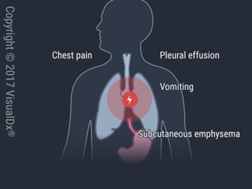
A 68-year-old man presented to the emergency department with chronic complaints of dysphagia and vomiting after meals, that developed 2 weeks prior. Hours earlier, he experienced a sudden onset of chest pain, vomiting, and hematemesis after a particularly heavy meal and binge drinking. The patient had a 25+ year history of alcohol use disorder. On physical examination, subcutaneous emphysema, tachycardia, and rebound tenderness were present. On imaging examination, a chest radiograph showed pneumothorax. What is the diagnosis?
Can you diagnose the patient? Use the Differential Builder in VisualDx to help you.
- AEsophageal motility disorder
- BThoracic aortic dissection
- CEosinophilic esophagitis
- DSpontaneous rupture of esophagus
Emergency Medicine Sample CME Question 3
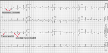
A 72-year-old woman presented to the emergency department with complaints of pleuritic chest pain, leg pain, and dyspnea that developed earlier that day. She had a medical history of chronic obstructive pulmonary disease (COPD) and pulmonary hypertension. On physical examination, tachycardia and unilateral leg swelling were present. ECG findings showed sinus tachycardia and an S1Q3T3 pattern of a prominent S wave in lead I, and presence of a Q wave and inverted T wave in lead III, indicative of right ventricular strain. What is the diagnosis?
Can you diagnose the patient? Use the Differential Builder in VisualDx to help you
- AAcute coronary syndrome
- BParoxysmal SVT
- CPneumothorax
- DPulmonary embolism
Emergency Medicine Sample CME Question 4

An 18-year-old woman presented to the emergency department with chest pain, dyspnea, and a cough that developed 4 weeks prior. The patient noted that these symptoms had gotten progressively worse over time. The patient admitted a chronic history of vaping cannabis and nicotine (5+ years). She had a medical history of generalized anxiety disorder and depression. A urine toxicology test was positive for THC. On physical examination, a fever of 38.5°C (101.3°F) was present. A chest CT showed diffuse patchy ground glass opacities in both bilateral upper and mid-lung zones. She developed hypoxia and nasal cannula oxygen requirements on the fourth day of hospitalization and was started on systemic steroids. She required noninvasive positive pressure but improved and was then extubated. What is the diagnosis?
Can you diagnose the patient? Use the Differential Builder in VisualDx to help you.
- AD. Viral pneumonia
- BB. Drug-induced pneumonitis
- CC. EVALI
- DA. Cryptogenic organizing pneumonia
Results
Provide your email to see your results and get exclusive discounts on additional board review questions.
VisualDX: Real-World Dermatology Cases + CME Credits
Comprehensive Dermatology Clinical Decision Support, specifically engineered for clinicians to visually improve decision making for optimal patient care. Features expert-curated, real-world case studies and guidance from practicing clinicians. Built by clinicians for clinicians.
Features:
-
Extensive library of dermatological conditions across diverse skin tones
-
Guided differential diagnosis tools for complex cases
- Clinical decision support at point-of-care
-
Educational resources for patient engagement
- Clinicans who use VisualDx save on average 2+ hours a week
CME Opportunities:
-
Earn up to 0.5 AMA PRA Category 1 Credits™ per search
-
CME credits automatically accrue through regular platform use
-
Joint accreditation through the Postgraduate Institute for Medicine
-
Credits available for physicians, pharmacists, and nurses
-
Access CME tracking and certificate printing through your personal account
Trusted by leading institutions and private practices, clinicians rely on VisualDX to help give accurate diagnoses, strengthen clinical decisions, and elevate patient care.
Your VisualDX subscription unlocks more than just diagnostic tools. Through joint accreditation with the Postgraduate Institute for Medicine, your VisualDx searches automatically earn up to 0.5 AMA PRA Category 1 Credits™. Access your account anywhere—from your institution’s network or via mobile—and watch your CME credits grow while enhancing your diagnostic accuracy.
“I had a patient who came for a consultation after being aggressively treated multiple times for a fungal infection that wasn’t improving. The previous diagnoses were incorrect, as the condition was actually pityriasis rosea. I used VisualDx to show the patient various photographs of the condition, which immediately reassured him and clarified the correct diagnosis.”
A.T., San Diego Family Dermatology Physician,